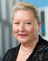
Ewa Snaar-Jagalska
Professor emeritus
- Name
- Prof. dr. B.E. Snaar-Jagalska
- Telephone
- 071 5272727
- b.e.snaar-jagalska@biology.leidenuniv.nl

I obtained my PhD at Leiden University (NL) in 1988 on G-protein and RAS mediated signal transduction in Dictyostelium. After a postdoctoral fellowship at the Johns Hopkins University, Medical School in Baltimore (USA) I returned to Leiden and was appointed as assistant professor in the workgroup Cell Biology and Genetics.
More information about Ewa Snaar-Jagalska
Postdocs
News
-
 Fighting gliobastoma brain tumours with two grants
 -
 Chemotherapy without side effects? It’s possible, with light
 -
 Leiden: The scene of the Zebrafish Disease Models conference 2018
 -
 Hunt for fundamental insight into and treatment for cancer
 -
 Ewa Snaar-Jagalska appointed as Professor in the field of cellular tumor biology
 -
 Aggressiveness of cancer cells halted
 -
 Completion of the Science Campus
 -
 New zebrafish study to understand human cancer
Former PhD candidates
I studied molecular signal transduction processes controlling chemotactic cell movements in the eukaryotic model Dictyostelium. I developed expertise in different aspects of signalling topics like G-protein coupled receptors (GPCRs), G-proteins, RAS, protein kinases and 14-3-3 proteins to answer the important open question: how can cells detect gradients of chemoattractants and translate this information into a chemotactic response? In 2000 I joined the Molecular Cell Biology section of IBL headed by Prof. H.P. Spaink, where I currently work as associate professor. In collaboration with Prof. T. Schmidt (Leiden Institute of Physics) we developed a new research line on the dynamic reorganization of plasma membrane interactions on single-molecule stimulation in vivo. This recently led to a breakthrough in the understanding of directional sensing of migrating cells. In 2003, I made a switch to a vertebrate model, the zebrafish. Currently my research focuses on the signalling dynamics underlying cancer and microenvironment interaction in zebrafish embryonic and transgenic models and in particular the role of innate immunity responses to cancer cells during angiogenesis and metastasis. In addition, I coordinate the ZF-CANCER project (2008-2011), an EU network for development of high-throughput bioassays for human cancers in zebrafish, with the ultimate aim of establishing zebrafish as a key vertebrate model for rapid, preclinical anticancer drug target discovery and lead compound selection. I also participate(d) in other previous (ZF-TOOLS, ZF-MODELS) and ongoing (ZF-HEALTH, SmartMix) international and national zebrafish research networks and co-organized several workshops on cancer and inflammation disease in zebrafish. The success of this new research line is shown by obtaining a medical grant for cancer research in the zebrafish model from the KIKA foundation (collaboration with several clinical research groups at the LUMC such as Molecular Cell Biology, Orthopedics, Pediatric Oncology and Pathology) as well as a NOW-TOP grant (collaboration with Prof. P. Hogendoorn (LUMC), Prof. T. Schmidt (LION) and Dr. A. H. Meijer (MCB-IBL)). I serve/served on the editorial board of several journals and am/was council member of various national and international organizations.
Research
I am interested in the signalling dynamics underlying cancer progression using zebrafish embryonic and transgenic models. The zebrafish is an excellent model system for this purpose: zebrafish form spontaneous tumors with similar histopathological and gene profiling features as human tumors; xeno-transplantation with human carcinoma cells is possible; and angiogenesis and immune response can be studied in vivo within the developing tumor.
The main focus of my research is on: directional cell migration and chemotaxis in cancer; the role of innate immunity responses to cancer cells during angiogenesis and metastasis; and human cancer modeling in zebrafish with the ultimate aim of establishing zebrafish as a key model for rapid discovery and assessment of cancer genetic targets (biomarkers) and drugs to develop strategies for cancer therapies. I closely collaborate with other groups at the Science Faculty and the LUMC and with other staff members of the Molecular Cell Biology group on development and application of high-throughput screening strategies for identifying novel anti-cancer drugs and cancer gene targets (with Prof. Herman Spaink) and on interaction between cancer and the immune system (with Dr. Annemarie Meijer). The research is funded by ZF-CANCER (EU FP7), ZF-HEALTH (EU FP7), SmartMix, KIKA foundation and NWO-TOP.
I have three main research themes:
Directional cell migration and chemotaxis in cancer
Cell migration is involved in vital physiological processes including embryogenesis and the immune response. However, it is likewise closely related to pathological conditions such as in cancer and chronic inflammatory responses. In many cases such cell migration is directional as determined by extracellular gradients of chemokines. Chemotaxis, the process of cell migration in chemokine gradients, is divided into the process of detection of the chemical gradient, the initial gradient sensing, and the subsequent translation of this gradient information into processes leading to physical movement and cell motility. Currently, little is known about the molecular mechanisms that control chemokine gradient sensing and migration of immune, endothelial and tumor cells. Fortunately, the molecular mechanisms that regulate these fundamental aspects of chemotaxis appear to be evolutionarily conserved and studies in the lower eukaryotic model system Dictyostelium discoideum in collaboration with Prof. Dr. Thomas Schmidt from Leiden Institute of the Physics have allowed us to form novel concepts, uncover molecular components, develop new single-molecule techniques, and test models of chemotaxis over the past years. In our earlier studies we found that a graded response in receptor mobility within the membrane, a distinct physical amplification mechanism towards downstream G proteins, and a high degree of membrane organization into signaling platforms does finally lead to a faithful signaling cascade and ultimately towards directed cellular motility.
Importantly, chemokines and chemokine receptors direct the migration of leukocytes to sites of inflammation and control leukocyte infiltration in cancer. In addition, chemokines also affect tumor growth by their angiogenic or angiostatic activity. Angiogenesis, the formation of new blood vessels from existing ones, is important in tumorigenesis to provide oxygen and nutrients and to stimulate the process of metastasis. Tumor growth occurs when the equilibrium between angiogenic and angiostatic factors is disturbed in favor of the angiogenic factors. The balance between angiostatic and angiogenic chemokines and their receptors expressed on the endothelial cell layer has to be strictly regulated. How this balance is regulated is still elusive.
In the current multidisciplinary project (NWO-TOP) we want to further develop, explore, and experimentally test concepts and molecular components of chemokine gradient sensing that leads to migration of immune, endothelial and tumor cells during tumor progression and angiogenesis in Ewing’s sarcoma using in vitro and zebrafish cancer models. The project aims at a molecular/mechanistic view of gradient sensing in tumor development. The fundamental knowledge generated in the course of the project has potential for application in anti-tumor immunity and anti-angiogenetic therapies for cancer treatment.
Group members involved: Sandra de Keijzer (PHD student, NWO-CW), Claudia Tulotta (PHD student, NOW-TOP) and Wietske van der Ent (PHD student on collaborative project with LUMC, funded by KIKA).
Collaborations: Prof. Pancras Hogendoorn (LUMC), Prof. Dr. Thomas Schmidt (LION) Dr. Annemarie Meijer (MCB-IBL), Prof. Herman Spaink (MCB-IBL).
Microenvironmental regulation of cancer angiogenesis and metastasis
How tumor cells interact with their microenvironment during tumor progression is a critical question in cancer biology. Answering this question requires live imaging of tumor-microenvironment interactions at the cellular level – a process severely limited in current animal models. To overcome this limitation, we established a xenograft model by injecting tumor cells into the blood circulation of transparent zebrafish embryos. This reproducibly results in rapid simultaneous formation of a localized tumor and micrometastasis, allowing time-resolved imaging at single-cell resolution. Roles of myeloid cells in critical tumorigenesis steps such as vascularization and invasion were revealed by genetic and pharmaceutical approaches. We discovered that the physiological migration of neutrophils controlled tumor invasion by conditioning the collagen matrix and forming the metastatic niche. Administration of VEGFR inhibitors enhanced migration of neutrophils, which in turn promoted tumor invasion. This work demonstrates the in vivo cooperativeness between VEGF signaling and myeloid cells in metastasis and provides a new mechanism underlying the recent findings that VEGFR targeting can promote tumor invasiveness (see figure above).
Currently we also study the role of immune cells in different aspects of cancer progression using novel transgenic lines with fluorescently marked immune cell population to facilitate real-time imaging. We attempt to unravel novel mechanisms of cancer inflammation and angiogenesis for the development of novel anti-tumor therapies based on targeting of vasculature and tumor-associated myeloid cells.
Group members involved: Shuning He (postdoc, ZF-CANCER project), Wietske van der Ent (PHD student on collaborative project with LUMC, funded by KIKA), Chao Cui (PHD student, SmartMix project) and Claudia Tulotta (PHD student, NOW-TOP).
Collaborations: Dr. Annemarie Meijer (MCB-IBL), Prof. Herman Spaink (MCB-IBL), Prof. Pancras Hogendoorn, Prof. Peter ten Dijke (LUMC), Drs. Jan-Willem Beenakker (PHD student LION), Drs. Veerander Ghotra (PHD student LACDR, ZF-CANCER) and Dr. Erik Danen (LACDR).
Human cancer modeling in zebrafish
Investigation of tumor migration and metastatic mechanisms are technically demanding (whole animal imaging) and expensive (instrumentation, animals). Replacement of small animals with a Danio rerio (zebrafish) embryo model is an alternative for these experiments. Because of the availability of transgenics, fluorescent reporter lines for vascular system and immune cells, and optical transparency, zebrafish is an excellent vertebrate model that allows the simultaneous in vivo imaging of cancer progression hallmarks. Recently we developed zebrafish xeno-transplantation assays to monitor cancer cell proliferation, migration, immune response, angiogenesis and metastasis formation within one week. The visual, non-invasive monitoring of cancer cells in transparent host embryos coupled with RNA interference and screens with chemical compounds enables the identification of novel gene targets and new compounds relevant for human cancer therapy, with the potential for commercial development. The zebrafish provides a fast, sensitive in vivo vertebrate model for identifying novel mechanisms of cancer progression and for development of medium to high-throughput application in preclinical target discovery and drug lead identification in a time- and cost-effective manner.
Group members involved: Shuning He (postdoc, ZF-CANCER project), Hanan Rian (PHD student, ZF-CANCER and ZF-HEALTH), Wietske van der Ent (PHD student on collaborative project with LUMC, funded by KIKA) and Claudia Tulotta (PHD student, NOW-TOP).
Collaborations: Prof. Herman Spaink (MCB-IBL), Prof. Pancras Hogendoorn (LUMC), Prof. Peter ten Dijke (LUMC), Prof. Clemens Lowik (LUMC), Drs. Veerander Ghotra (PHD student LACDR, ZF-CANCER) and Dr. Erik Danen (LACDR).
Professor emeritus
- Faculty of Science
- IBL
- Animal Sciences
The staff member publications service is not available
The staff member activities service is not available
- geen


