

Free online course teaches you all about imaging
How do you microscopically image zebrafish larvae? The new free online course ‘Imaging the Vertebrate embryo’ teaches you all about it. Leiden PhD candidates Salomé Muñoz Sánchez and Radoslaw Gora contributed to the course.
Massive Open Online Course, or MOOC. That is how this popular type of online course is called, and millions of people worldwide make eager and free use of it. MOOCs come in all shapes and sizes and cover just about every imaginable subject. And now, there is also one about imaging: the microscopic visualisation of biological material, specifically the embryos of vertebrates.
Investigating tuberculosis
The course is part of the European project ImageInLife, established in 2017 to exchange knowledge about imaging and to train a new batch of top scientists. Researchers in Leiden have been using imaging techniques to study infectious diseases, especially tuberculosis, for years, says Annemarie Meijer, professor of Immunobiology and training coordinator of ImageInLife. ‘The transparent larvae of the zebrafish are exceptionally well suited for this.’
Sharing knowledge
And now, it is time to share the know-how from ImageInLife with the outside world. ‘In this MOOC, you will learn how to image vertebrate embryos and how to subsequently process the data and images,’ it says on the website. The MOOC focuses primarily on zebrafish and mice.
Leiden contribution
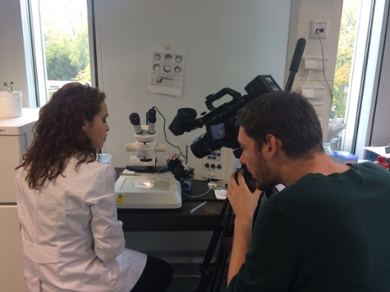
PhD candidate Salomé Muñoz Sánchez from Meijer's group is part of the team behind the MOOC. In the course, she shows how you can inject zebrafish to study human diseases. ‘We demonstrate the injection of pathogenic bacteria into the blood or tissue of zebrafish larvae. This allows you to visualise the interactions between bacteria and immune cells in real-time.’
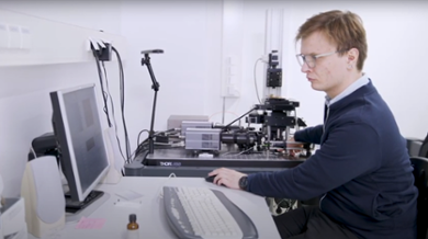
Radoslaw Gora is also part of the MOOC. The PhD candidate from Marcel Schaaf's group discusses the technique single-molecule microscopy (SMM). ‘In the course, we show how to prepare a zebrafish embryo for research. We then demonstrate how to image the zebrafish embryo using a type of SMM called Total Internal Reflection Fluorescence (TIRF) Microscopy.'
The course starts on 20 April and ends on 15 June. Students spend about 2 hours per week on the MOOC. More info can be found here.