
This is how a channel is formed between two organelles
The channel through which two cell components exchange material appears to form at the edge of their contact surface, and not in the middle. This was discovered by the Leiden physical chemists Jelger Risselada and Edgar Blokhuis together with researchers from the University of Lausanne in Switzerland. They published their findings in The Journal of Physical Chemistry Letters on 20 February.
The beginning: a striking observation
It all starts with a remarkable observation in Lausanne. Swiss researchers examine so-called vacuoles of yeast cells under the microscope to look for channels between them. These vacuoles are not very interesting in themselves, but they resemble human cell parts, namely the organelles. Because of this resemblance, they can provide interesting insights. Normally, the channels between vacuoles are too small to be seen under a normal microscope. But the researchers come up with a handy trick for this: with the help of osmotic pressure they manage to enlarge the vacuoles and their channels in such a way that the channels become visible.
And what they then see surprises them. The Swiss researchers are curious about the connecting channel between the two yeast ‘organelles’. For a cell to function properly, it is essential that organelles can exchange material with each other, for example, to discharge waste products. This transfer takes place via a channel. However, it was unknown where such a channel is located in the contact surface between two organelles. But then the researchers see something interesting through their microscope: the connecting channel appears to be at the edge, as can be seen on the right. Last author Jelger Risselada: ‘Then we wondered, is that always the case? Or only if you magnify these channels unnaturally, as the Swiss did?’
A Nobel Prize-worthy subject
Risselada decides to look for the answers and engages colleague Edgar Blokhuis. ‘Edgar is a specialist in the tricky mathematics needed for the models I wanted to develop,’ says Risselada.
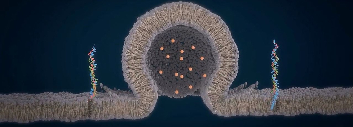
Research into channels, also called fusion pores, is hot. In 2013, for example, three American scientists received the Nobel Prize for their discovery of the so-called SNARE proteins. ‘These proteins bring two organelles together and also direct the formation of the channel,’ says Risselada. ‘In textbooks, channels are always reproduced symmetrically, in the case of neurons (see animation) but also in the case of the much larger organelles. What we have now discovered shows something completely different for organelles.’
Due to the selected cookie settings, we cannot show this video here.
Watch the video on the original website orThis animation shows how neurotransmitters in the brain are released in a synapse, the connection between two neurons, which are nerve cells in the brain. The SNARE proteins provide a symmetrical connection between a so-called vesicle filled with neurotransmitters and the membrane of a neuron so that the neurotransmitters can flow into the synapse.
Credits: Je-Kyung Ryu
How the channel really forms
Because organelles are large, two organelles that exchange material have a large contact surface. This means that a channel can be created in several places. Risselada: ‘That’s why a channel does not have to be neatly in the middle of the contact surface, but can also sit on the edge.’ In order to get a definite answer, Risselada first performs molecular simulations. Next, Blokhuis starts working with a theoretical model to explain the outcome of the simulations mathematically. ‘Our calculations show that the channel is always located at the edge. This can also be seen in our animation,’ says Risselada.
That animation shows how a channel that is initially in the middle moves to the edge of the contact surface. In this way the channel loses its symmetry: the membrane of the channel is almost straight on one side and very curved on the other side. ‘You would think that every system strives for symmetry, but this asymmetry turns out to be the most optimal situation energetically speaking.’ This also has consequences for the canal, Blokhuis says. ‘Our model shows that the canal increases in size when it is on the edge. That’s what biology wants.’
Simulation of a channel
Due to the selected cookie settings, we cannot show this video here.
Watch the video on the original website orThis simulation by the researchers shows that the channel between the organelles is located at the edge. The membranes of the two organelles are shown in white, which have now merged and between which a channel has formed.
Influencing the channel size from the outside
And that wasn’t the only insight the Leiden chemists came up with. ‘We also saw that the size of the channel depends on the angle of contact between the two organelles, which is equal to the attraction of these organelles,’ explains Risselada. ‘And you can increase that attraction by adding certain proteins. So we have also discovered a mechanism that shows that external proteins can influence the channel size between two organelles,’ explains Risselada. ‘This finding differs from recent findings published in the journal Nature (Bai et al, 2018), in which the authors actually suggest that the growth of the channel requires direct integration of SNARE proteins.’
Publication
Edgar M. Blokhuis, Massimo D’Agostino, Andreas Mayer and H. Jelger Risselada. Fusion Pores Live on the Edge (2020). The Journal of Physical Chemistry Letters
Text: Bryce Benda