
Novel detection method for iron in Alzheimer’s brain
For many years, scientists have observed a correlation between Alzheimer’s disease and a surplus of iron in the brain. However, a causal link between the two has not been proven yet. We lack knowledge concerning the specific form of iron that is involved in the development of neurodegenerative diseases. Now, a collaboration of physicists and medical researchers shows how they can detect and quantify some distinctive forms of iron in post-mortem brains. Publication on December 12 in Nature’s Scientific Reports.
Iron intake
We eat daily between ten and twenty milligrams of iron. After being absorbed by the gut, this metal participates in a wide variety of essential metabolic processes, including oxygen transportation, DNA replication and electron transport. However, if it’s not properly regulated, iron can cause damage to cells.
MRI
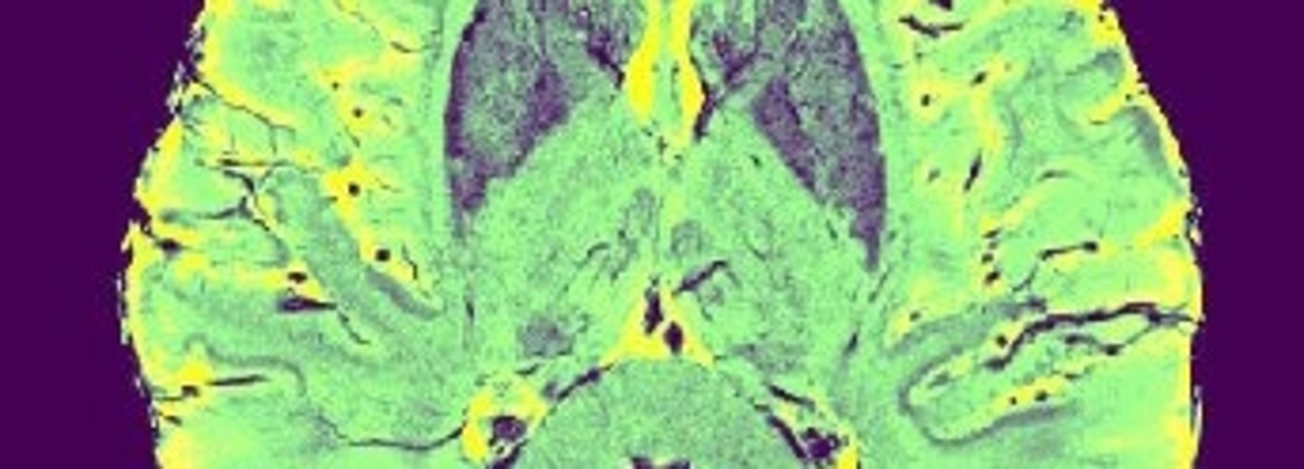
In the brains of patients with neurodegenerative diseases, such as Alzheimer’s and Parkinson’s disease, iron is upregulated: it can increase to up to three times greater than the normal level. Scientists can use MRI scans to indirectly image iron in post-mortem brain tissue, but the data interpretation remains much debated. Such measurements are indirect, affected by several sources of artifacts, and poorly quantitative.
Magnetism and biology
A multidisciplinary team of Leiden physicists (LION) and radiologists from the Leiden University Medical Centre (LUMC), led by postdoc Lucia Bossoni, has developed a method based on the combination of Electron Paramagnetic Resonance Spectroscopy (EPR) and SQUID magnetometry to detect and quantify complementary species of iron in post-mortem brain tissue. In combination with MRI, this approach will allow scientists to better understand the role played by iron in Alzheimer’s disease.
Publication
Pravin Kumar, Marjolein Bulk, Andrew Webb, Louise van der Weerd, Tjerk H. Oosterkamp, Martina Huber, and Lucia Bossoni, ‘A novel approach to quantify different iron forms in ex-vivo human brain tissue’, Nature’s Scientific Reports
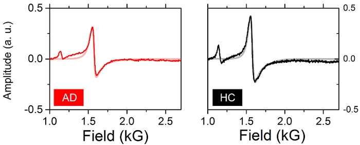
X-band EPR spectra of brain tissue. The graphs show a comparison between Alzheimer’s disease tissue (left) and healthy control tissue (right). The potentially toxic iron appears as a band at a magnetic field of about 1.5 kG.
Credit header image: M. Buijs