Research project
New generation of graphene biosensors based on smooth surfaces and sharp edges
The surface and the edges of graphene are expected to provide higher sensitivity and specificity in detecting and characterizing single molecules. However fundamental physical limits exist in reaching an ultimate precision in detecting the dynamics of chemical and biological systems. The research in the group of Dr. Gregory Schneider focuses on two fundamental aspects that need to be characterized in order to use graphene as an ideal sensor material: i) how to effectively interface graphene devices with biological materials so that detection becomes sensitive and selective, and ii) understand and characterize the chemical reactivity of ‘just made’ graphene edges. Exploiting the full potential offered by graphene as a natural material in sensing applications will only be possible through in-depth fundamental research of these two limiting aspects.
- Contact
- Grégory Schneider

There is still a lot to learn about graphene. Physics and engineering aspects of this material are booming, but the most simple aspects of chemistry, for example a simple chemical reaction, is still difficult to quantify with this material. If the fundamental chemistry of this material has been very poorly investigated to date, it is primarily because graphene is not an 'easy' material to work with.
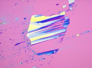
What is graphene? The purest graphene is the one obtained from mother nature: a single flake of exfoliated graphene. Think about this flake floating in air where edges and surfaces are available for a chemical game! Every chemist will argue that multitude of opportunities exist, but unfortunately, graphene in air does not exist. Main research focus is to overcome the physical complexity of graphene, so that edges and surface become readily accessible for 'a' chemical game: the edges and the two surfaces will start communicating for the purpose of biosensing. To do so, the unique edge and surface chemistry of graphene will be harvested for developing a general method for functionalizing graphene through a fundamental understanding of graphene chemistry at a single molecule level.
Pioneering work on graphene single molecule sensors
DNA sequencing is very rapidly growing into an industry of major interest. A variety of techniques exist, each with their own pros and cons. The use of nanopores – nanoscale holes in a membrane – for DNA sequencing was proposed more than 20 years ago. The idea is straightforward: pass a DNA molecule through the pore from head to tail, and read off each base when it is located at the narrowest constriction of the pore, using the ion current passing through the pore as the probe for detecting the identity of the base. While biological pores were investigated for quite some time, solid-state nanopores are now emerging. They have tunable pore size, are more stable than biological membranes, offer re-usability upon cleaning, and allow for scaling and device integration. But DNA sequencing has so far not been demonstrated with these devices : conventional silicon-based nanopore membranes are relatively thick, typically ~30 nm, which corresponds to ~60 bases along a single-stranded DNA molecule. While solid-state nanopores are excellent new tools for biophysical studies, they are therefore not directly useful as-is in DNA sequencing applications. Recently however, graphene nanopores were introduced. Graphene forms the ultimate nanopore membrane since it is a carbon sheet with a thickness of only a single atom. Furthermore it is electrically conductive, which opens up new modalities of measuring the traversing nucleotides, for example by running a tunneling current through the DNA molecule that is traversing a graphene gap, to directly probe the chemical nature of the bases. To these ends, however, the chemistry of graphene (at the edge & on the surface) must be first fundamentally understood, preferably down to a single molecule level. Our group is currently taking this challenge.
Exploiting the full potential offered by graphene in sensing applications requires extensive fundamental studies of the behaviour of the surface and of the edges of graphene upon their interaction with biological systems (lipids, proteins, enzymes, DNA, RNA and ultimately biological cells) as well as a quantification of the measurable electronic response of the graphene surface (and edge respectively) caused by a biological stimuli such as the presence (respectively the passage) of a biomolecule. The surface and the edges of graphene operate as sensors in two fundamentally different directions: in a typical solution-gated graphene field-effect transistor, the surface is sensitive to charge transfer conferred by a molecule in the vicinity of graphene and therefore can detect a single molecule as a whole, while edges can be used as atomically flat electrodes that could transversally sense the precise structure and chemical composition of a biomolecule passing close to the edges. In both cases, biomolecules are being sensed, but the level of output information is different: surfaces can trap, detect and sense while edges can provide sequence information. This holds the potential that one can combine both and use the surface to selectively trap and identify, guide electrophoretically the trapped molecule towards the edge (using for example a mobile lipid bilayer) and obtain sequence information using a transverse electrochemical current generated between two edges separated by a physical gap on the order of the lateral dimension of the biomolecule (i.e., the backbone of an unfolded protein or DNA, typically < 5nm).
Surface
Graphene (in a field-effect transistor device) is highly sensitive to the interaction of molecules at its surface as the binding of molecules alters the carrier density through electrostatic gating and charge transfer, resulting in changes in the direct current (DC) conductance of the sensor. Furthermore, graphene is hydrophobic and therefore not compatible with most hydrophilic biological elements. Our group research focuses on two key challenges for developing biologically-relevant graphene-based chemical and biosensors are i) to develop a graphene sensor platform that is less impeded by unfolding or denaturing factors (in a device that can also operate as a charge sensor), and ii) to develop a hybrid (but not covalently modified) graphene interface that is biocompatible, selective, reversible, sensitive and that will allow specifically the detection of biologically and disease relevant proteins.
Edges
The two key challenges for the development of new graphene sensing technology exploiting the atomically sharp edges of sheet-like graphene are i) to define, control and characterize the chemistry of graphene edges, and ii) to tailor (electro)chemically their functionalization for their subsequent use as active electrodes. Prior to defining chemistry, developing protocols for the controlled fabrication of reactive graphene edge-states (zig-zag and armchair) is essential. In fact, similarly to the surface structure of normal catalysts, the precise edge structure of graphene is key to its chemical reactivity: graphene with zig-zag versus armchair edges make an enormous difference to the pi-electron system and hence to their chemical interaction with co-reactants.
Where we expect our research to head in the near future
The first outcomes of our research will be to validate that graphene field effect transistors can be used at their full sensing power (without intrinsic physical limitations arising from physiological conditions or poor control of the most simple chemical aspects involved in sensing). If so, graphene will be a powerful material to detect single molecules. In medicine/biology, for most proteins, researchers have no methods to measure or detect them (besides indirectly using DNA or RNA). Even when a good and specific antibody is available researchers often need too much sample to get a signal strong enough to reach a conclusion. Ultrapure, defect-free, high-mobility graphene has the potential for reaching ultimate sensitivity, as it was demonstrated for the detection of individual gas molecules. Biodetection, however, needs more sophisticated strategies and importantly devices that are properly interfaced with biological environments without suffering from consequent limitations. If such a device would exist (similar as a gas sensor) then it would allow that only a few molecules of a specific protein in blood (or other body fluids) could be detected: a clear jump forward in diagnostics and medicine.
Our research links several disciplines, and also addresses fundamental questions about very simple concepts such as chemistry of C-H edges (or other functionality) on graphene, preservative atom-by-atom sculpting of graphene, and more importantly the combination of both: how to chemically control and functionalize an atomically thin edge of graphene to either specifically sense one biomolecule or controllably scan a sequence of individual molecules. It also addresses other important scientific questions on device fabrication at a level of a few carbon atoms (while standard lithographic techniques barely have a resolution of a few tens of nanometers at minimum), understanding how the current density in graphene nanoribbons and gaps scale with molecular dimensions of the fabricated structure, what is the impact of specific chemistry of graphene edges and surface on the conductance and tunnelling/electrochemical current of the structure made, what is the chemistry required to prevent Pi-stacking interactions known to induce unspecific interaction of DNA and proteinic materials with graphene, chirality of the functional graphene edges and their impact on a chiral separation of molecules, to name a few.
Key publications
- Schneider, G.F., Q. Xu, S. Hage, S. Luik, J.N.H. Spoor, S. Malladi, H. Zandbergen, C. Dekker, "Tailoring the hydrophobicity of graphene for its use as nanopores for DNA translocation", Nature Communications, vol. 4, Oct, 2013. DOI: 10.1038/Ncomms3619
- Schneider, G.F., C. Dekker, "DNA sequencing with nanopores", Nature Biotechnology, vol. 30, no. 4, pp. 326-328, Apr, 2012. DOI: 10.1038/Nbt.2181
- Schneider, G.F., V.E. Calado, H. Zandbergen, L.M.K. Vandersypen, C. Dekker, "Wedging Transfer of Nanostructures", Nano Letters, vol. 10, no. 5, pp. 1912-1916, May, 2010. DOI: 10.1021/Nl1008037
- Schneider, G.F., S.W. Kowalczyk, V.E. Calado, G. Pandraud, H.W. Zandbergen, L.M.K. Vandersypen, C. Dekker, "DNA Translocation through Graphene Nanopores", Nano Letters, vol. 10, no. 8, pp. 3163-3167, Aug, 2010. DOI: 10.1021/Nl102069z
