

Hooray! This extremely sensitive microscope survived its relocation
Moving an electron microscope of 2000 kg is a delicate challenge. The highly sensitive instrument needed to be moved to a new measurement hall, but even a tiny bump could damage it. After a few nerve-racking weeks of preparing the move and reinstalment, the researchers finally have a verdict: the instrument still works.
The instrument in question is a low-energy electron microscope (LEEM) that had been standing in the same place for 13 years. The move is such a complicated task that it had been in the works for months.
A nerve-racking move
To get the job done, a team of many specialists was involved. Not only was the move itself plotted out in detail, but the new location was also prepped to carry the weight and have all the provisions the researchers need. And that’s not to mention labelling every cable to ensure it’s reinstalled correctly in the new location. Even two technicians from the German company that produced the microscope came over to help.
Physicist Amin Moradi reflects on the day of the move: ‘It was incredibly tricky because the smallest bump could cause damage and delay the work for months or even break it beyond repair.’ Inside the LEEM, a beam of electrons is redirected several times through various components. If one of the components moves just a little bit, the beam is misaligned and the microscope does not work. Repairing this would be a tedious process but luckily the team is spared the work, says Moradi: ‘After two weeks of reinstalling the LEEM, we turned it on and it immediately worked!’
-

A group of expert technicians, researchers and movers is required to get the job done. -
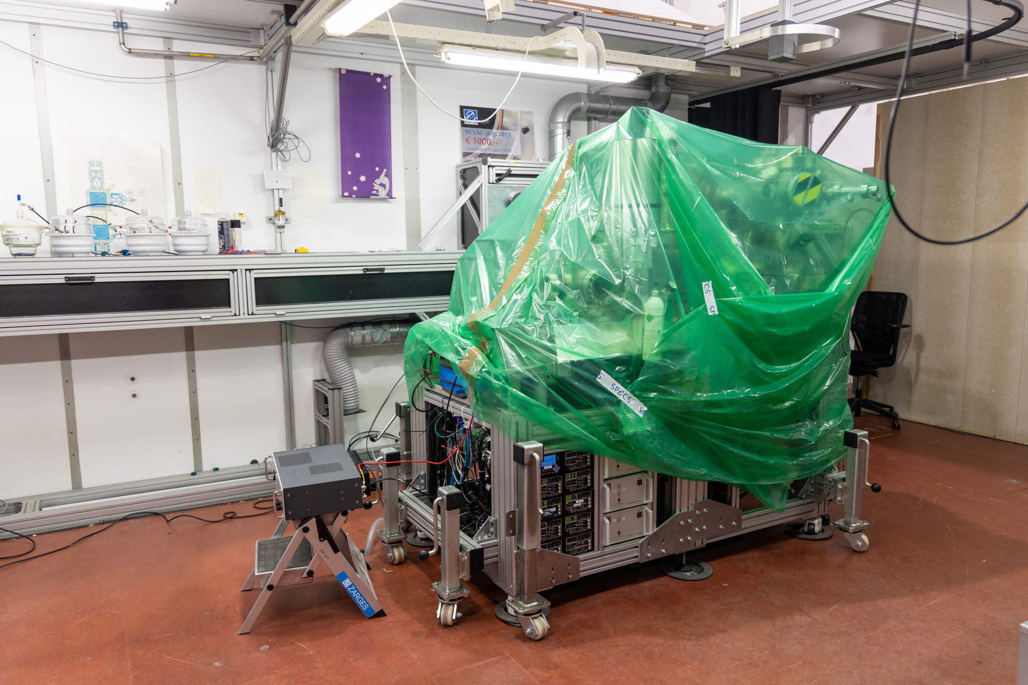
The microscope is all wrapped up and placed on wheels in the old measurement hall, ready to be moved. -

The smallest bump can damage the instrument, so extreme care is required. -
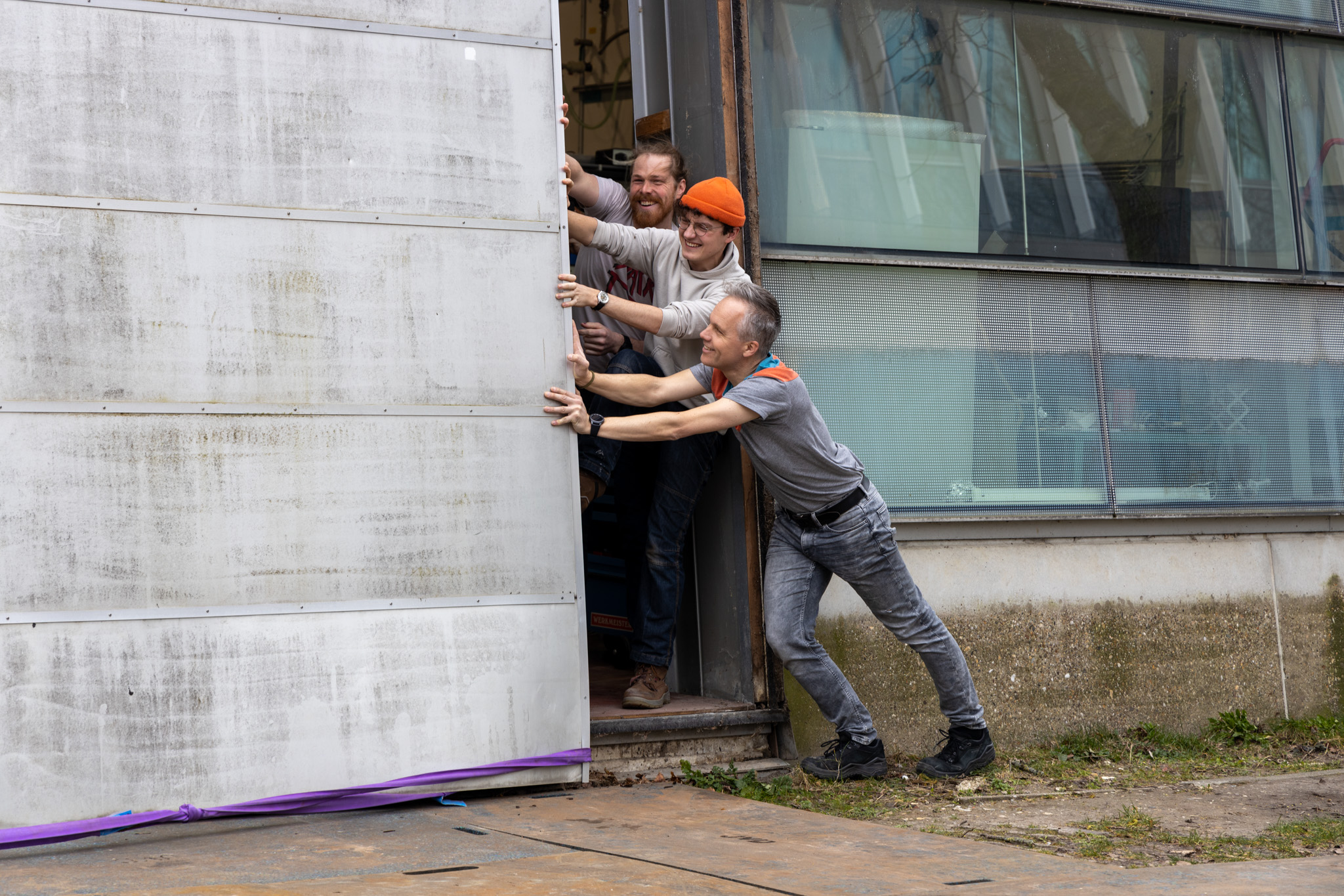
An unexpected setback: the door of the old measurement hall won’t open fully. -

Lifting the extremely sensitive instrument to a truck, because it is too large to move through the building. -
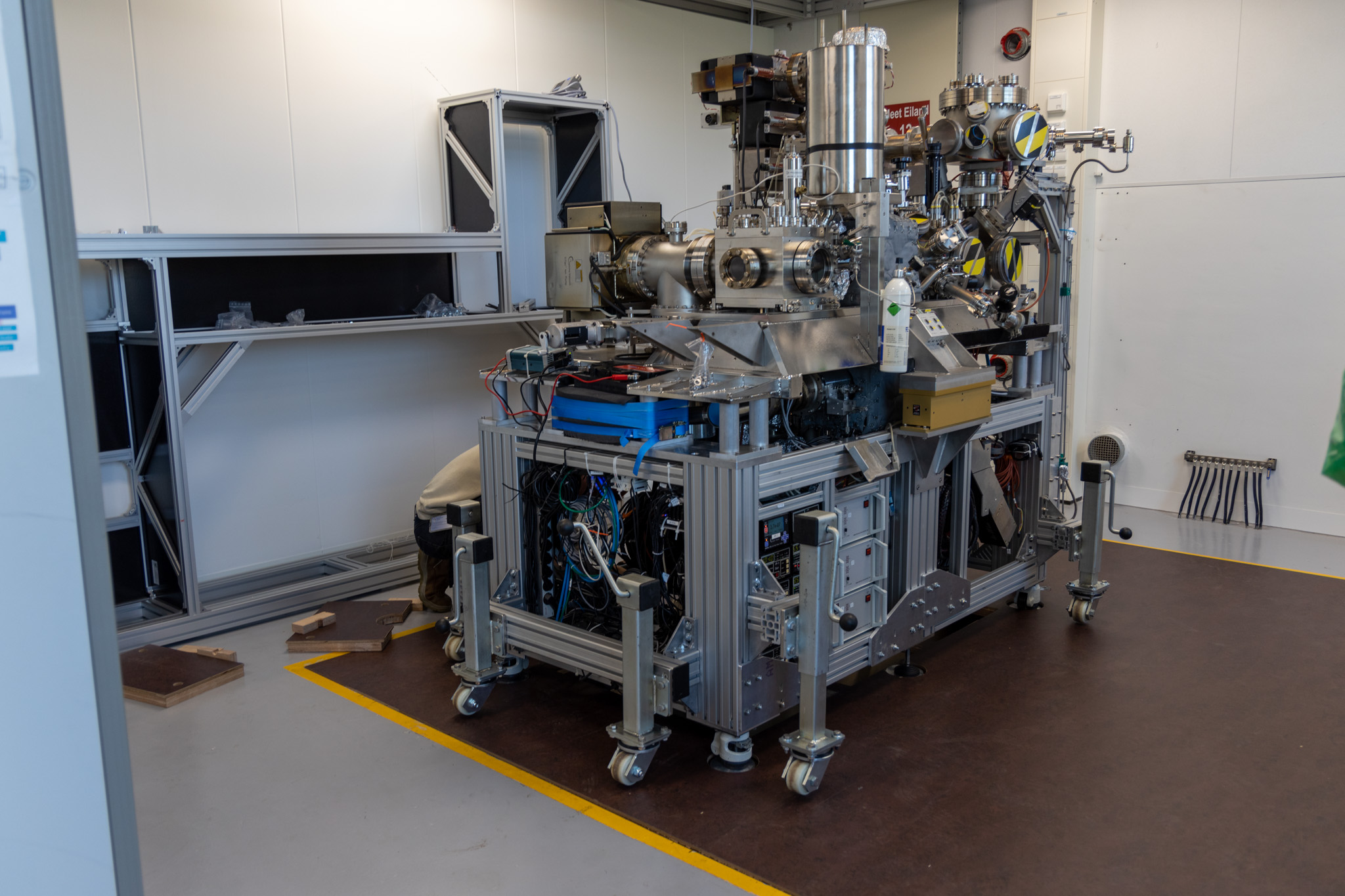
The LEEM made it safely to its new location.

For what research is this immense instrument used?
The LEEM has a better resolution than an optical microscope that uses photons, but there is one downside: the sample has to be in a vacuum. And for biological samples like human cells, a vacuum changes their structure. ‘For a long time, we’ve had this dream to combine the techniques of an optical and electron microscope’, says Moradi. ‘Because then we can study such samples when they are emerged in water, wíth nanometer resolution.’ That dream is now close to becoming reality, as Moradi and his colleagues are adding the optical component to the LEEM.
With this add-on, he creates an optical near-field electron microscope (ONEM) inside the LEEM that combines the benefits of both an optical and electron microscope. Moradi visualises the new technique with Indonesian shadow puppetry.
‘The puppets are backlit behind a screen so that the audience only sees a shadow. In the case of ONEM, the puppet is the sample and the screen a photocathode. Photons come from the light source, pass the sample and are turned into electrons when they hit the photocathode. The sample is placed extremely close to the photocathode to achieve a high resolution. Comparing again to the puppetry: if the puppet is close to the screen, you see a very clear shadow. But if it is placed further away, the shadow becomes blurry.’
Moradi took his first image to prove the technique works, but then his work was interrupted by the move. ‘I can’t wait to continue to see what is possible, we might be the first to pull this off!’