

Tracers that light up tumours help surgeons
How do surgeons avoid causing nerve damage or leaving cancerous cells behind? An interdisciplinary research group at the LUMC hopes to improve operations and make them less invasive with the aid imaging techniques. They are working with medical companies to make these techniques widely available.
This is an article in a series about partnerships and societal impact.
If patients need to have a tumour removed, they go for a scan at the radiology department to establish the precise location of the tumour. A contrast liquid is often used to identify where it is. But the scan can only be viewed on a computer, which isn’t much use to the surgeon during the operation. ‘Once the surgeon is operating he can’t see a clear difference between healthy tissue and tumour as he can on the scan,’ explains LUMC researcher Fijs van Leeuwen. ‘Our joint goal is for surgeons to see more, for instance by having tumour cells light up.’
Making diseased cells visible
Since 2006 the LUMC has been focusing on various imaging technologies that can be used in operations. This is known as image-guided surgery. One such technology uses tracers that bind to tumours. These can be radioactive, fluorescent or a combination of the two, and can be seen clearly in the body. ‘The tracers bind to the malignant cells, but disappear from the rest of the body, making the diseased areas much more visible for the surgeon,’ says LUMC surgeon Alexander Vahrmeijer. The techniques are mainly used to remove tumours. ‘It’s crucial that no single tumour cell is left behind. Otherwise, the disease can return,’ says Van Leeuwen. ‘But there are other applications, such as detecting bacterial infection on prostheses or lighting up nerves that mustn’t be damaged during operations.’
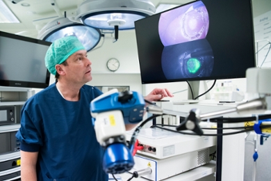
The signal from the tracers usually can’t be seen with the naked eye. Special detection techniques are needed, for instance a camera that can detect fluorescence together with a regular film camera. Surgeons then have live images in the operating theatre of the tissue that they are operating on and where in this tissue the tracers can be found. Vahrmeijer: ‘We’ve spent the last few years developing some of these techniques with businesses such as Quest Medical Imaging or Karl Storz Endoscopy.’
Google Maps for operation
A second imaging technique in surgery uses augmented and/or virtual reality. Radiological scans can be projected over the patient during an operation, thus creating a kind of Google Map of the site of the tumour. The technique brings together radiological imaging before an operation and surgical imaging in the operating theatre. For this research the researchers from the LUMC also worked with various partners from industry. Their findings can be used outside oncology too, in orthopaedics and neurosurgery, for instance.
Routine care
A number of techniques have been developed with industry that are now used in clinical care. These vary from commercially available tracers that were developed by the LUMC to detection techniques such as cameras sold worldwide for image-guided surgery.
‘At the LUMC and other centres we routinely use fluorescent tracers when operating on colon cancer that has spread to the liver and in sentinel lymph node procedures with head and neck tumours,’ says Vahrmeijer. ‘But most image-guided techniques are not used in routine care. They are only used in patients as part of research.’ Van Leeuwen adds, ‘By working with market leaders in surgical techniques, we can ensure that people all around the world benefit from our research. That is what motivates me to work with industry.’
Photo above: Fijs van Leeuwen (l) and Alexander Vahrmeijer with one of the cameras for image-guided surgery that they are developing together with industry. (Photo: LUMC)
How fluorescent tracers help surgeons remove tumours
Due to the selected cookie settings, we cannot show this video here.
Watch the video on the original website or