

Resolution of electron microscope greatly improved
The use of a new type of detector has generated a strong improvement in the resolution of electron microscopes. The 'low-energy electron microscope' (LEEM) can now be used for research on the thinnest possible materials. The tests with the detector represent the first result of the ESCHER project.
Greater sensitivity
Research into the development and characteristics of advanced materials can now be carried out more efficiently and with greater sensitivity. The resolution of the LEEM can even be improved by a factor of 2.5. The tests have been carried out by scientists in Leiden, Twente and IBM.
Low energy electron microscope
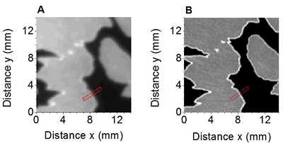
Low energy electron microscopy is a recent development within the field of microscopy. The technique is becoming increasingly important in research into processes in micro- and nanoelectronics, such as semiconductor nano-crystal growth, graphene synthesis and other phenomenons at nanoscale. ‘In a conventional electron microscope, electrons are accelerated to high energies to irradiate the sample,' says Ruud Tromp, Professor of the Physics of Surface Materials, and member of the research staff of IBM. What's special about LEEM is that it uses low-energy electrons. This makes the electrons very sensitive to even the finest structures on the surface. Magnetic electron lenses use the reflected electrons to form a video image of the sample and even of its electronic properties. Of course, a better detector results in a better image.'
Medipix2
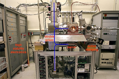
The new detector, the Medipix2, is a high-tech CMOS detector developed by the Medipix2 consortium. The primary aim of Medipix2 is to detect X-rays. Researchers Sense Jan van der Molen, Irakli Sikharulidze and Ruud Tromp installed the Medipix2 in Raoul van Gastel's LEEM instrument at the University of Twente.
Night-vision cameras
The detector that has been used to date is the same type as that used in night-vision cameras. Unfortunately, it has many shortcomings. It can handle only a limited signal sensitivity, the signal measured suffers from noise, and the spatial resolution is limited. The Medipix2 resolves all three of these problems in one fell swoop. Given the excellent results, the researchers expect that Medipix2 will be standard in future LEEM instruments.
Graphene
The advantages of the improved resolution are significant for the development of micro- and nanotechnology. It will now become possible to study the development and the characteristics of graphene in greater depth. Graphene is a thin material just a single atom thick. It is well known that graphene opens up new possibilities in the field of electronics and spintronics, for which Albert Fert and Peter Grünberg were awarded the Nobel Prize in 2007.
Protein molecules
How can graphene be produced efficiently? For the time being, the important step from laboratory to industry has not yet been made. The growth of graphene can be observed live in a LEEM. This can provide insights into how the material can be made in the future on a large scale. The microscope also signifies a step forward for research into the exotic combination between metals and organic materials. For ESCHER, a series of experiments is planned with self-assembled monolayers, organic layers of exactly one molecule in thickness. By introducing such layers and choosing the right molecules, a surface takes on new characteristics, such as waterproofing of even its opposite of water permeability.
ESCHER microscope
The ESCHER microscope will be installed at Leiden University later this year. The instrument, based on a design by Ruud Tromp, will be further developed by the team in Leiden. With the new detector, the microsope's resolution and contrast will be greatly improved. The improved microscope will then be able to handle studies at temperatures varying from close to absolute zero to above 1500 degrees celsius.
This research has been part-funded by the Netherlands Organisation for Scientific Research (NWO) and the STW Technology Foundation. The Medipix2 consortium is co-ordinated by CERN and NIKHEF.
(7 July 2009)