
Images
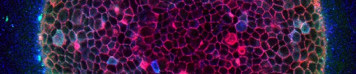
Confocal image of an epithelial cyst cultured in 3D collagen gel
By Leo Price.
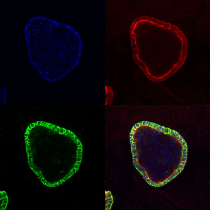
The cyst was originally seeded as a single cell. After 12 days of proliferation, polarization of the epithelial cells and subsequent apotosis of luminal cells, a cyst with a single-cell thick wall results. The cyst is labeled with rhodamine phalloidin to label f-actin (red), DAPI (blue) and anti-E-cadherin antibodies (green).