
Images
Confocal image from a primary mouse mammary epithelial cell cyst
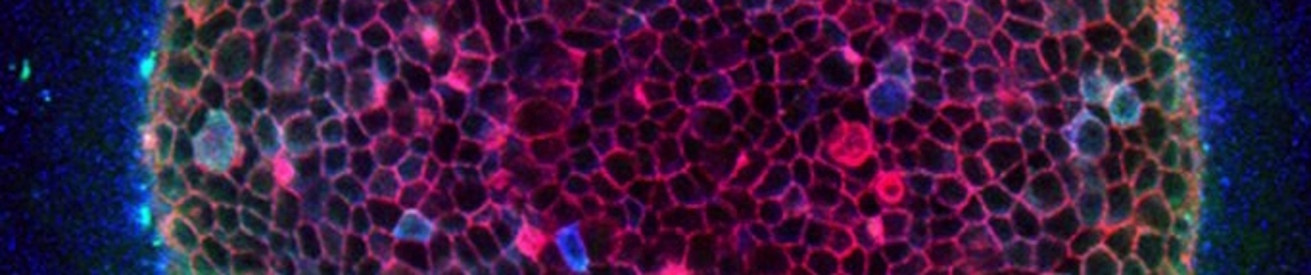
Matrigel reconstitutes the in vivo environment of the mammary gland. Mammary epithelial cysts plated in Matrigel form a lumen due to the lack of matrix attachment of the luminal cells. Approximately 4-5 days after plating the cells start to branch.
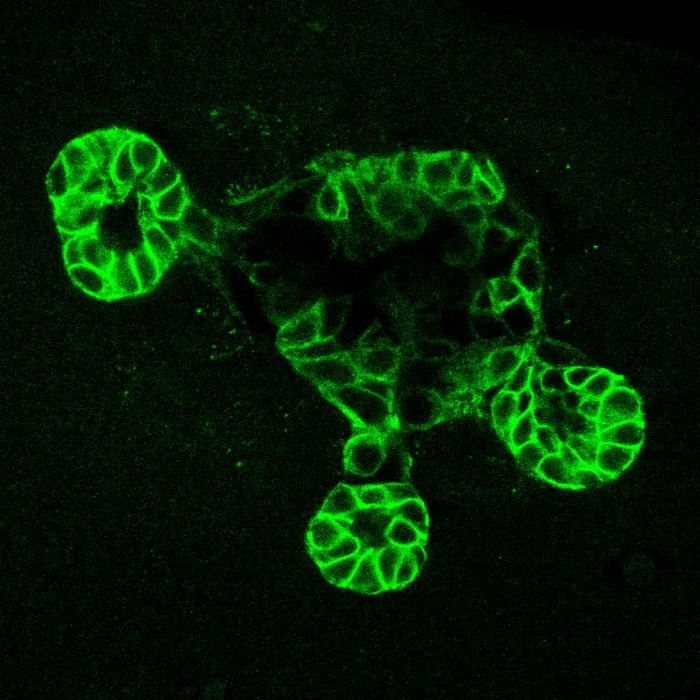
This is a confocal image from a primary mouse mammary epithelial cell cyst that was plated in Matrigel.