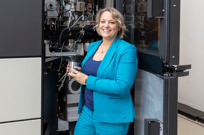
Ariane Briegel: 'AI literally opens new worlds for the life sciences'
Bacteria caught red-handed, deeply frozen just as they were about to cause Lyme’s disease. Ariane Briegel is wildly enthusiastic about the wonders she observes thanks to three elements: a freezing technique, a camera-equipped microscope, and AI. ‘It’s fascinating. Every single cell is different.’
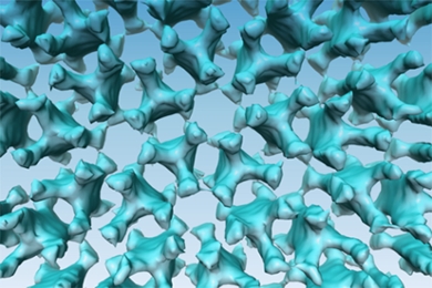
Bacteria follow their ‘nose’ towards a food source. Well, strictly speaking not their nose, but their receptor proteins. Neatly ordered like a honeycomb, Briegel has watched them prick through cell walls. On the outside they catch signals; on the inside they set the cell to work swimming in the right direction.
From 100 slices to 3D images with smart software
The camera in Briegel’s microscope takes approximately one hundred photographs of the same number of cell slices. Smart software then turns these hundred photographs into an understandable 3D image. This has literally opened a new world for Briegel and her colleagues: they can observe in great detail what cell structures and organs look like and how bacteria ‘get moving’.
And the fun has only just begun
And the fun has only just begun. ‘When I completed my PhD twenty years ago, I was producing two datasets a day at most. These days it’s forty to fifty a day, far more than researchers could ever look at. And we want to tackle even larger projects.’ Like mapping the 1.5 kilograms of bacteria on and inside the human body. To this end, Briegel works with AI researchers to develop learning algorithms that recognise individual cell components and incorporate them separately in the images. This is currently done by human experts, but will soon be fully automated.
Infection, cancer, or growth: we can map it all
At the same time, together with other labs, Briegel’s group works on further developing microscopy technology: ‘We can already see larger structures, fixed in plastic. The complete intestines of zebrafish, for example. The camera in the microscope takes pictures day and night for a whole week, and we use AI to try and identify where interaction with cholera bacteria takes place. This analysis is where our challenge lies now, which is why we collaborate with AI experts. Infection, cancer, growth: with these techniques, we will soon be able to map in detail so many biological systems in the life sciences.
Text: Rianne Lindhout
Photo: Patricia Nauta