
Research project
Oncogenic protein tyrosine kinases and Annexin family members in breast cancer formation and metastasis
Researcher: Marjo de Graauw
- Contact
- Marjo de Graauw
The progression of breast cancer depends, in part, on the ability of tumor cells to invade and metastasize. This metastatic spread is thought to mimic the cell migration that occurs during embryogenesis and is initiated by a phenotypic switch from an epithelial to mesenchymal phenotype (Fig. 1). This so-called EMT enables cells to acquire the ability to execute the multiple steps of the invasion–metastasis cascade. During an EMT, epithelial cells lose their cell–cell interactions and cell polarity, and undergo a major change in their actin cytoskeleton. This enables them to acquire a mesenchymal appearance with increased motility and invasiveness
Phosphotyrosine proteomic screening revealed phosphorylation of the lipid-, calcium-, and actin-binding protein, annexin A2 (AnxA2) at Tyr23 as a major event preceding ts-v-Src kinase-induced cell scattering and EMT. We have shown that phosphorylation of AnxA2 mediates cell scattering and branching morphogenesis (Fig. 2) in vitro via regulation of the actin severing protein cofilin (de Graauw et al. MCB Feb 2008). Current research focuses on the role of (phosphorylated) AnxA2 and its closed family member AnxA1 in breast cancer formation and metastasis. Different breast cancer cell lines are used for our studies, including MTLn3 (highly invasive rat breast carcinoma), MCF7 (low invasive human breast carcinoma) and MDA-MB-231 (highly invasive, human breast carcinoma). In these cells both AnxA2 and AnxA1 are localized in actin rich membrane protrusions that are formed during migration, suggesting that both proteins are involved in regulating the actin cytoskeletal network during migration and invasion. To understand their precise role in cancer, we use different approaches, which include, human breast cancer tissue arrays (> 500 patients) (Fig. 3), in vivo models, live cell imaging to study random cell migration and invasion and 3D morphogenesis assays. For more information on annexins click on the following link.
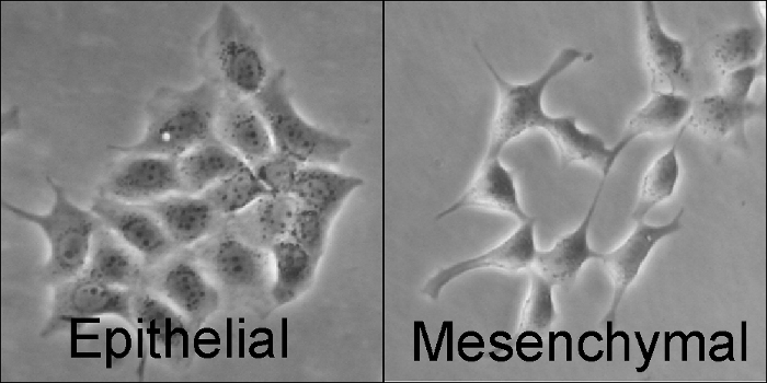
Figure 1: Activation of Src kinase results in an EMT. Cells expressing an inactive Src tyrosine kinase form tight cell-cell interaction and obtain an epithelial phenotype (left). Upon phosphorylation/activation of Src kinase, cells change their actin cytoskeletal network, loose cell-cell interactions and obtain a migratory, mesenchymal phenotype (right).
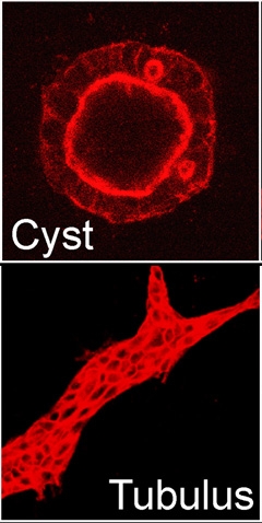
Figure 2: Phosphorylation of AnxA2 results in spontaneous tubulogenesis. Cells expressing either WT-AnxA2 (left) or a phospho-mimicking Y23E-AnxA2 (right) were grown in collagen gels for 6 days. WT-AnxA2-expressing cells developed into cysts, while cells expressing Y23E-AnxA2 developed into tubules, a process that normally only occurs in the presence of the growth factor HGF.
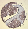
Figure 3: AnxA2 expression in human breast cancer. Human breast cancer samples were spotted on a tissue array and stained for AnxA2. The AnxA2 protein is highly expressed in tumor cells, while stroma surrounding the cancer cells is negative.