
New method of detecting rapid virus development
Physicists, including Leiden researcher, Sanli Faes, have devised a new technique for studying processes at microscale rapidly and extremely precisely. This new method will make it easier to develop antiviral medication. And it doesn’t stop there. Publication in ACS Nano.
Viruses spread effectively
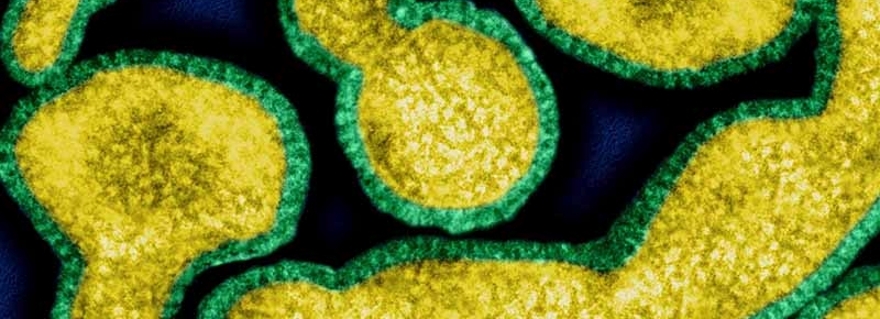
Using this new technique, researchers can actually see viruses grow. Viruses, such as influenza, spread very effectively and can therefore be fatal for their host. They are able to multiply spontaneously, at a very fast rate and in huge numbers. Not only this, they are also minuscule.
Preventing viruses
Once researchers understand how viruses are formed, they may be able to develop a preventive medicine that will stop them occurring. To do this, a system is needed that can track viruses at nanometre scale with a time resolution of less than a millisecond. Leiden physicist Sanli Faez, along with researchers from Harvard and Jena, MIT, the Leibniz Institute of Photonic Technology and glass fibre manufacturer Heraeus Quarzglas, has developed such a technique. The international team, led by Harvard University, has published the results in ACS Nano with Faez as the lead author.
Challenges
The two challenges in mapping the formation of viruses are speed and size. Fluorescence microscopy can detect individual proteins, but this technique is too slow to properly determine the process. It is like trying to take a photo of the wing movements of a hummingbird using a standard camera: parts of the process can be recorded but crucial frames are missing.
Very small particles, such as protein shells, can be observed by the way in which they disperse light. This technique, known as elastic scattering, emits a limitless number of photons, which resolves the speed problem. But the photons interact with dust particles that partly obscure the particles in which the researchers are interested.
Glass fibres
The team of researchers decided to make use of glass fibre technology; the exceptional quality of these fibres has been perfected over recent years by the telecoms industry. The researchers designed a new glass fibre cable that contains a nanoscale channel, smaller than the wavelength of light. The channel is filled with a liquid containing nanoparticles. When light is transmitted through the channel, the nanoparticles scatter the photons, which the researchers are able to capture using a microscope. This method has enabled the researchers to determine the movement of viruses with a diameter of 26 nanometres at a speed of thousands of measurements per second.
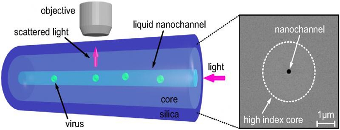
The method of studying processes at nanoscale.
Versatile technique
‘The main goal of our research was to develop a general tracking technique based on elastic scattering to overcome the limitations of fluorescence microscopy,’ Faez explained. ‘We worked with this virus to demonstrate the system, but as well as research on viruses, there are many other possible applications, such as counting the number of vesicles in bodily fluids. Cancer cells produce more vesicles than healthy cells, so if we can measure the number of vesicles precisely enough, we may be able to develop a new diagnostic method for cancer. Screening on the basis of electron microscopy is too expensive for this.’
Viruses in new light (Video)
Sanli Faez et al. Fast, Label-Free Tracking of Single Viruses and Weakly Scattering Nanoparticles in a Nanofluidic Optical Fiber, ACS Nano, October 27, 2015